Artificial intelligence (AI) continues to revolutionize every field—even cancer research, where it is showing immense potential to overcome many of the hurdles that hinder timely and accurate cancer diagnosis and treatment. In under-resourced settings, for example, clinicians face limited access to new technology, expert pathologists, and other important resources, which means many patients aren’t diagnosed with cancer until it’s too late. And even in well-resourced environments, the subjective nature of some analyses can lead to missed or delayed diagnoses, underscoring the need for more consistent and accurate diagnostic tests.
At the AACR Annual Meeting 2025 in Chicago, researchers discussed the capacity of AI to help overcome some of these logistical challenges during the “Artificial Intelligence and Machine Learning for Basic and Translational Research” Minisymposium.
“This session highlights really exciting research showing how AI and [machine learning] can leverage multiple modes of data to improve different kinds of predictions, from diagnoses, subtyping of cancers, targeted therapies, and prognoses,” said Samantha Riesenfeld, PhD, of the University of Chicago, who moderated the session together with Paul Spellman, PhD, of UCLA Health Jonsson Comprehensive Cancer Center. “Several talks also show how critical information can be better extracted from more accessible types of patient samples, such as blood samples and pathology images.”
During the session, held April 27, researchers explored a wide range of AI applications in cancer research, including AI-driven models designed to analyze routine clinical data to diagnose either cancer cachexia or skin cancer with high accuracy.
Predicting Cancer Cachexia Through AI
In one presentation, Sabeen Ahmed, a graduate student at the University of South Florida and Moffitt Cancer Center, reported on an AI-driven model that analyzes routinely collected clinical data to predict a patient’s likelihood of developing the debilitating symptom known as cancer cachexia, which is characterized by systemic inflammation, severe muscle wasting, and profound weight loss.
While cancer cachexia can be a side effect of any cancer type, it frequently afflicts patients with pancreatic cancer, impacting not only their quality of life but also their ability to enroll in clinical trials and undergo certain cancer treatments. Cancer cachexia is estimated to cause about 30% of all cancer-related deaths, and researchers continue to explore how to manage this serious complication, as well as how to better catch cachexia before it’s too advanced.
“Detection of cancer cachexia enables lifestyle and pharmacological interventions that can help slow muscle wasting, improve metabolic function, and enhance the patient’s quality of life,” said Ahmed. “Unfortunately, current methods for detecting cancer cachexia rely on clinical observations, weight loss thresholds, and indirect biomarkers, which are often inconsistent, subjective, and detected too late in disease progression.”
To improve the detection of cancer cachexia, Ahmed and colleagues developed an AI model, called SMAART-AI (for Skeletal Muscle Assessment Automated and Reliable Tool based on AI), that integrates imaging scans and multiple types of routine clinical data to report the probability that a patient has or will develop cancer cachexia. The computed probability is an example of an AI-driven biomarker, which is a measurable indicator of cancer cachexia identified or derived through machine learning, Ahmed noted.
“Compared with traditional biomarkers, AI-driven biomarkers may enable more sensitive and accurate detection of cancer cachexia by uncovering complex patterns across multiple data modalities that may be missed by traditional analyses,” she said.
SMAART-AI works through two main steps. In the first step, it examines diagnostic images, such as computed tomography (CT) scans, to quantify the amount of skeletal muscle in the patient’s body using an algorithm that automatically detects and measures muscle. In a validation test, the AI model’s quantifications of skeletal muscle differed by a median of 2.48% from the manual quantifications made by expert radiologists.
“On average, the model’s measurements of skeletal muscle were very close to the expert radiologists’ measurements, demonstrating the high reliability of our AI-based approach,” said Ahmed. “Our framework also provides a model confidence estimate, which helps flag outputs that are likely to deviate significantly from manual assessments, thus enabling more informed interpretation and potential human review when necessary.”
In the second step, SMAART-AI integrates the skeletal muscle quantification with multiple types of clinical data routinely collected as part of a cancer diagnosis workup—lab results, notes from electronic medical records, and weight and height measurements, among others. Ahmed explained that a key innovation of the study was the use of large language models to process unstructured clinical notes to extract relevant information that may not be captured in structured lab reports or imaging results.
“Each type of data gives a different clue about whether a patient has cancer cachexia, but individually, they might not tell the full story,” Ahmed noted. “By combining all these data modalities, our AI-driven biomarker model is designed to identify hidden patterns and make a more accurate diagnosis of cancer cachexia than any single test alone.”
Ahmed reported that when AI-driven skeletal muscle quantification was combined with information about tumor stage and patient demographics, weight, and height, the model accurately identified cachexia in 77% of pancreatic cancer cases. This accuracy increased to 81% with the addition of lab results and further to 85% when clinical notes were incorporated.
“Our AI-driven approach provides a scalable and objective solution for detecting cancer cachexia using data collected at the time of cancer diagnosis, potentially allowing health care providers to initiate interventions to mitigate cachexia earlier in the disease course,” said Ahmed. “The findings highlight the growing potential of machine learning to revolutionize cancer care and enable personalized treatment plans.”
Using AI-driven Analysis of Pathology Images to Diagnose Skin Cancer
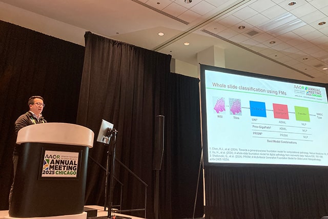
In another presentation, Steven Song, an MD/PhD candidate at Pritzker School of Medicine and the Department of Computer Science at the University of Chicago, discussed how pretrained AI-based models could help diagnose nonmelanoma skin cancer (NMSC), potentially even in resource-limited settings like parts of Bangladesh, where individuals have an increased risk of NMSC due to high levels of arsenic in contaminated drinking water.
As Song explained, skin lesions suspected of being NMSC are typically resected, thinly sliced, and mounted on a slide for evaluation by a pathologist, but in many regions around the world, the lack of expert pathologists limits the ability to review slides and diagnose NMSC quickly.
While researchers have often proposed AI-based tools to fill in these resource gaps, Song noted that these types of tools are usually custom-made and require resources not readily available in many places, including computational experts, specialized computational hardware, and large amounts of data upon which to train AI models.

As an alternative, Song and colleagues explored the potential of “off-the-shelf” AI tools to guide NMSC diagnosis. One strategy is to pretrain models on vast amounts of data in resource-rich environments before deploying them in settings with limited access to large datasets and/or the specialized equipment or experts needed for developing models from scratch, Song noted.
In his presentation, he reported the accuracy of three pretrained models—PRISM, UNI, and Prov-GigaPath—in identifying NMSC from digital pathology images of suspected cancerous skin lesions. All three models work by converting a high-resolution digital image of a tissue pathology slide into small image tiles, extracting meaningful features from the tiles, and analyzing these features to compute the probability that the tissue contains NMSC.
The models’ accuracy in diagnosing NMSC was evaluated on 2,130 tissue slide images representing more than 500 biopsy samples from Bangladeshi individuals enrolled in the Bangladesh Vitamin E and Selenium Trial. Of the 2,130 total images, 706 were of normal tissue, and 1,424 were of confirmed NMSC.
Accuracy of the three models was compared with that of ResNet18, an established but older architecture for image recognition. “ResNet architectures have been used as a starting point for training vision models for nearly a decade and serve as a meaningful baseline comparison for evaluating the performance gains of newer pretrained foundation models,” Song explained.
PRISM, UNI, and Prov-GigaPath each significantly outperformed ResNet18—correctly distinguishing between NMSC and normal tissue in 92.5% (PRISM), 91.3% (UNI), and 90.8% (Prov-GigaPath) of cases, compared with an accuracy of 80.5% for ResNet18.
To make the pretrained models more amenable to use in resource-limited settings, Song and colleagues developed and tested simplified versions of each model. The simplified models, which require less extensive analysis of pathology image data, still significantly outperformed ResNet18, with accuracies of 88.2% (PRISM), 86.5% (UNI), and 85.5% (Prov-GigaPath), demonstrating robustness even with reduced complexity, according to the researchers.
“Overall, our results demonstrate that pretrained machine learning models have the potential to aid diagnosis of NMSC, which might be particularly beneficial in resource-limited settings,” said Song.
He noted, however, that “further work is needed to address practical considerations, such as the availability of digital pathology infrastructure, internet connectivity, integration into clinical workflows, and user training.”
Learn more about AI’s applications in cancer research and care:
- Experts Forecast Cancer Research and Treatment Advances in 2025: In an interview with Cancer Research Catalyst, the official AACR Blog, Regina Barzilay, PhD, discussed AI’s potential roles in cancer risk prediction, drug development, multimodal data analysis, and treatment selection.
- Interfacing with the Future: Artificial Intelligence in Oncology: In a Plenary Session during last year’s AACR Annual Meeting, researchers examined the potential of AI to assess medical images, address global health disparities, match patients with targeted therapies, and identify therapeutic vulnerabilities.


