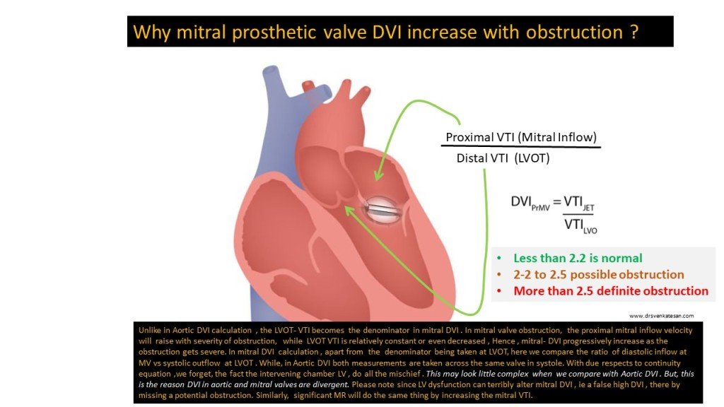
Why prosthetic mitral valve DVI increase with obstruction, while Aortic valve DVI, decreases?
December 23, 2024 by dr s venkatesan

Final message
Prosthetic valve assessment is complex, thought process intensive examination. Not every echocardiographer can do it efficiently. It needs a good knowledge of anatomy, physiology of inter & Intra valvular hemodynamics .It demands thorough understanding of principles of Doppler echocardiography and also the hidden truths( ie, How we take liberty with the mighty Bernoulli equation for granted )
In spite of the number of imaging and doppler parameters we are able to gather ,still, we need to analyze them with reference to the clinical presentation. Mind you, even an innocuous episode of fever, associated dyspnea, and tachycardia can elevate the mitral gradient and sound a false alarm.
Depending solely on prosthetic valve gradients to diagnose obstruction is the biggest error we commit. We have seen this, even from elite hospitals. Echocardiography is not the final say, one may require cine fluoroscopy, CT scan or even PET in appropriate situations.
Reference


1.Zoghbi WA, Jone PN, Chamsi-Pasha MA, Chen T, Collins KA, Desai MY, Grayburn P, Groves DW, Hahn RT, Little SH, Kruse E, Sanborn D, Shah SB, Sugeng L, Swaminathan M, Thaden J, Thavendiranathan P, Tsang W, Weir-McCall JR, Gill E. Guidelines for the Evaluation of Prosthetic Valve Function With Cardiovascular Imaging: A Report From the American Society of Echocardiography Developed in Collaboration With the Society for Cardiovascular Magnetic Resonance and the Society of Cardiovascular Computed Tomography. J Am Soc Echocardiogr. 2024 Jan;37(1):2-63. doi: 10.1016/j.echo.2023.10.004. PMID: 38182282.
2.H, Freeman WK. Echocardiographic Assessment of Prosthetic Valves. Rev Cardiovasc Med. 2022 Oct 11;23(10):343. doi: 10.31083/j.rcm2310343. PMID: 39077122; PMCID: PMC11267339.


